
Page 213 - 磁共振成像2024年7期电子刊
P. 213
综 述||Reviews 磁共振成像 2024年7月第15卷第7期 Chin J Magn Reson Imaging, Jul, 2024, Vol. 15, No. 7
mrTRG 评 分 系 统 的 准 确 性(72.9% vs. 38.1%;P< 表2 基于T2WI联合DWI的mrTRG标准
0.001)和 阅 片 者 间 一 致 性(k=0.580 vs. 0.338;P< Tab. 2 The mrTRG Criteria Based on Combined T2WI
and DWI
0.001)均优于原有的基于 T2WI 序列的 5 级 mrTRG 分级 类别 T2WI+DWI影像特征
评分系统,且改良三级 mrTRG 与 NCRT 术后 3 年无 mrTRG 1 完全缓解 信号正常,T2WI和DWI无肿瘤证据。
病生存期独立相关。然而,目前尚缺乏系统性研究 mrTRG 2 近似完全缓解 DWI少许高信号区,T2WI显著低信号纤维瘢
痕区。
对比这三种评分系统的优劣,这需要进一步探讨和 mrTRG 3 中等缓解 T2WI低信号区超过DWI高信号残余灶。
验证。 mrTRG 4 轻微缓解 DWI高信号残余灶超过T2WI低信号区。
mrTRG 5 无缓解 瘤灶表现与初始状态一致,无纤维化证据。
1.3 基于DCE-MRI序列的mrTRG评分系统研究现状
注:DWI为扩散加权成像;mrTRG为磁共振肿瘤退缩分级。
动态对比增强 MRI(dynamic contrast-enhanced
MRI, DCE-MRI)是一种利用对比剂来增强图像的 血管外细胞外空间体积比等在评估肿瘤的血流动力
MRI技术。DCE-MRI的定量参数,如容量转移常数、 学特性方面发挥重要作用,可能有助于确定对 NCRT
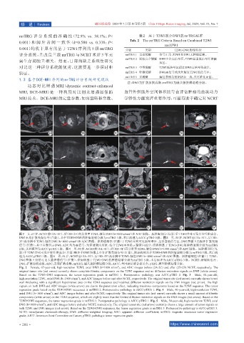
图 2 女,67岁,NCRT前(2A~2C)、后(2D~2F)高分辨率 T2WI、轴位 DWI(b=1000 s/mm²)和 ADC图像。原肿瘤部位(红箭)于 T2WI序列可见完全纤维成分、
DWI未见扩散受限信号(白箭),基于 T2WI+DWI的肿瘤退缩分级为 mrTRG 1级,术后病理为 AJCC-pTRG 0级。图 3 男,50岁,NCRT 前(3A~3C)、后(3D~
3F)高分辨率 T2WI、轴位 DWI(b=1000 s/mm²)和 ADC 图像。原肿瘤部位(红箭)于 T2WI 序列可见肠壁增厚、呈显著低信号区,DWI 图像上局限性扩散受限
信号(红箭),其中白箭所示 DWI、ADC 均为高信号,为穿透效应所致,结合 T2WI 序列提示黏蛋白成分,该病例基于 T2WI+DWI 的肿瘤退缩分级为 mrTRG
2
2级,术后病理为 AJCC-pTRG 1级。图 4 男,40岁,NCRT 前(4A~4C)、后(4D~4F)高分辨率 T2WI、轴位 DWI(b=1000 s/mm )和 ADC 图像。原肿瘤部位(红
箭)于 T2WI序列可见少量纤维成分(白箭)略多于 DWI图像上少许扩散受限信号(红箭),则该病例基于 T2WI+DWI的肿瘤退缩分级为 mrTRG 3 级,术后病
2
理为 AJCC-pTRG 1级。图 5 男,54岁,NCRT前(5A~5C)、后(5D~5F)高分辨率 T2WI、轴位 DWI(b=1000 s/mm )和 ADC图像。原肿瘤部位(红箭)于 T2WI、
DWI 图像上仍然可见大量肿瘤信号(红箭),该病例基于 T2WI+DWI 的肿瘤退缩分级为 mrTRG 5 级,术后病理为 AJCC-pTRG 3 级。NCRT:新辅助治疗;
DWI:扩散加权成像;ADC:表观扩散系数;mrTRG:磁共振肿瘤退缩分级;AJCC:美国癌症联合委员会;pTRG:病理肿瘤退缩分级。
Fig. 2 Female, 67-year-old, high-resolution T2WI, axial DWI (b=1000 s/mm ), and ADC images before (2A-2C) and after (2D-2F) NCRT, respectively. The
2
original tumor site (red arrow) currently shows complete fibrotic components on the T2WI sequence and no diffusion restriction signals on DWI (white arrow).
Based on the T2WI+DWI sequences, the tumor regression grade is mrTRG 1. Postoperative pathology was AJCC-pTRG 0. Fig. 3 Male, 50-year-old,
2
high-resolution T2WI, axial DWI (b=1000 s/mm ), and ADC images before and after NCRT, respectively. The original tumor site (red arrow) currently shows bowel
wall thickening with a significant hypointense area on the T2WI sequence and localized diffusion restriction signals on the DWI images (red arrow). The high
signals on both DWI and ADC images (white arrow) are due to the penetration effect, indicating mucinous components based on the T2WI sequence. The tumor
regression grade based on the T2WI+DWI sequences is mrTRG 2. Postoperative pathology is AJCC-pTRG 1. Fig. 4 Male, 40-year-old, high-resolution T2WI,
axial DWI (b=1000 s/mm ), and ADC images before and after NCRT, respectively. The original tumor site (red arrow) currently shows a small amount of fibrotic
2
components (white arrow) on the T2WI sequence, which are slightly more than the limited diffusion restriction signals on the DWI images (red arrow). Based on the
T2WI+DWI sequences, the tumor regression grade is mrTRG 3. Postoperative pathology is AJCC-pTRG 1. Fig. 5 Male, 54-year-old, high-resolution T2WI, axial
DWI (b=1000 s/mm ), and ADC images before and after NCRT, respectively. The original tumor site (red arrow) currently shows a large amount of tumor signals on
2
both T2WI and DWI images (red arrow). Based on the T2WI+DWI sequences, the tumor regression grade is mrTRG 5. Postoperative pathology is AJCC-pTRG 3.
NCRT: neoadjuvant chemoradiotherapy; DWI: diffusion-weighted imaging; ADC: apparent diffusion coefficient; mrTRG: magnetic resonance tumor regression
grade; AJCC: American Joint Committee on Cancer; pTRG: pathologic tumor regression grade.
·206 · https://www.chinesemri.com