
Page 109 - 磁共振成像2024年7期电子刊
P. 109
临床研究||Clinical Articles 磁共振成像 2024年7月第15卷第7期 Chin J Magn Reson Imaging, Jul, 2024, Vol. 15, No. 7
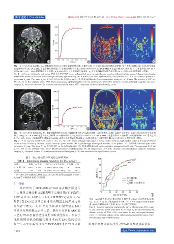
图 2 女,53岁,TAO活动期。2A:脂肪抑制 T2WI示左侧上睑提肌复合体、双侧下直肌、外直肌及内直肌肌腹明显增粗,信号增高(绿箭);2B:时间-信号强度
曲线为平台型;2C:DCE-MRI定量参数V 伪彩图,左/右侧眼外肌V 均值分别为0.994/0.959;2D:DCE-MRI半定量参数AUC伪彩图,左/右侧眼外肌AUC最大
e e
值为4.847/3.362。TAO:甲状腺相关性眼病;DCE-MRI:动态对比增强磁共振成像;V :血管外细胞外间隙容积分数;AUC:时间-信号强度曲线下面积。
e
Fig. 2 A 53-year-old female with active TAO. 2A: FS-T2WI shows enlarged left superior rectus-levator complex, bilateral inferior rectus, bilateral lateral rectus
and bilateral medial rectus with increased signal intensity (green arrow); 2B: A plateau-type time-signal intensity curve pattern; 2C: DCE-MRI-derived quantitative
parameters V map. The mean V are 0.994/0.959 on the left/right orbit; 2D: DCE-MRI-derived semi-quantitative parameters AUC map. The maximum AUC are
e
e
4.847/3.362 on the left/right orbit. TAO: thyroid-associated ophthalmopathy; FS: fat suppression; DCE-MRI: dynamic contrast-enhanced magnetic resonance
imaging; V : fractional volume of the extravascular-extracellular space; AUC: area under the time-signal intensity curve.
e
图3 女,56岁,TAO非活动期。3A:脂肪抑制T2WI示右眼上睑提肌复合体、内直肌及双侧下直肌稍增粗,双侧下直肌信号稍增高(绿箭);3B:时间-信号强度曲
线为平台型;3C:DCE-MRI定量参数V 伪彩图,左/右侧眼外肌V 均值分别为0.708/0.587;3D:DCE-MRI半定量参数AUC伪彩图,左/右侧眼外肌AUC最大值为
e
e
2.276/1.863。TAO:甲状腺相关性眼病;DCE-MRI:动态对比增强磁共振成像;V :血管外细胞外间隙容积分数;AUC:时间-信号强度曲线下面积。
e
Fig. 3 A 56-year-old female with inactive TAO. 3A: FS-T2WI shows enlarged right superior rectus-levator complex, right medial rectus and bilateral inferior
rectus without obviously increased signal intensity (green arrow); 3B: A plateau-type time-signal intensity curve pattern; 3C: DCE-MRI-derived quantitative
parameters V map. The mean V are 0.708/0.587 on the left/right orbit; 3D: DCE-MRI-derived semi-quantitative parameters AUC map. The maximum AUC are
e
e
2.276/1.863 on the left/right orbit. TAO: thyroid-associated ophthalmopathy; FS: fat suppression; DCE-MRI: dynamic contrast-enhanced magnetic resonance
imaging; V : fractional volume of the extravascular-extracellular space; AUC: area under the time-signal intensity curve.
e
表2 TAO活动性分期的独立影响因素
Tab. 2 Independent imaging predictors for TAO activity
参数 系数 标准差 比值比(95%置信区间) 瓦尔德 P值
AUC最大值 0.633 0.292 1.883 (1.063~3.336) 4.712 0.030
V 均值 2.745 1.113 15.570 (1.757~137.983) 6.082 0.014
e
注:TAO 为甲状腺相关性眼病;AUC 为时间-信号强度曲线下面积;
V 为血管外细胞外间隙容积分数。
e
3 讨论
本研究基于 DCE-MRI 对 TAO 患者眼外肌进行
半定量及定量分析,结果表明半定量参数(TTP均值、
AUC 最大值、AUC 均值)和定量参数(V 最大值、Ve
e
图 4 AUC 最大值、V 均值及其联合模型评估 TAO 活动性的 ROC 曲
e
均值)在 TAO 活动期组和非活动期组之间差异均具 线。AUC:时间-信号强度曲线下面积;V :血管外细胞外间隙容积分
e
数;TAO:甲状腺相关性眼病;ROC:受试者工作特征。
有统计学意义。其中,V 均值和 AUC 最大值是 TAO Fig. 4 Receiver-operating characteristic curves of maximum AUC, mean
e
活动性分期的独立影响因素。联合V 均值和AUC最 V and combined model (maximum AUC+mean V ) for discriminating the
e
e
e
disease activity of TAO. AUC: area under the the time-signal intensity
大值对 TAO 患者活动性分期诊断效能较高。相较于 curve;V : fractional volume of the extravascular-extracellular space; TAO:
e
thyroid-associated ophthalmopathy.
既往采用其他功能磁共振技术研究 TAO 临床活动
性 [28-29] ,本文创新性地应用 DCE-MRI 评估 TAO 患者 眼外肌微循环灌注改变,为TAO分期提供新依据。
·102 · https://www.chinesemri.com