
Page 90 - 磁共振成像2024年7期电子刊
P. 90
磁共振成像 2024年7月第15卷第7期 Chin J Magn Reson Imaging, Jul, 2024, Vol. 15, No. 7 临床研究||Clinical Articles
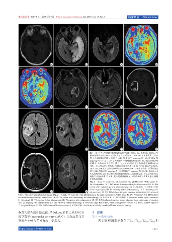
图1 男,47岁,左侧颞叶胶质母细胞瘤(WHO 4级)。1A:术前CE_T1WI示左
侧颞角强化病变;1B~1E 为同步放化疗后复查;1B:T2-FLAIR 术区见片状水
肿 ;1C:MUSE-DWI 呈高信号 ;1D:增强前 T1 mapping 图 ;1E:增强后 T1
mapping图;1F:CE_T1WI示左侧颞叶不规则强化病变;1G:随访强化灶较前明
显减小,为治疗相关改变。图 2 女,50 岁,右侧顶叶胶质母细胞瘤(WHO
4级)。2A:术前CE_T1WI示右侧顶叶强化病变;2B~2E为同步放化疗后复查;
2B:T2-FLAIR术区边缘高信号灶;2C:MUSE-DWI以低信号为主,周围环绕高
信号;2D:增强前T1 mapping图;2E:增强后T1 mapping图;2F:CE_T1WI示术
区边缘强化灶;2G:随访强化范围较前明显增大,为肿瘤复发。CE_T1WI:对比
增强 T1加权成像;FLAIR:液体衰减反转恢复;MUSE-DWI:多重灵敏度编码
扩散加权成像。
Fig. 1 Male, 47 years old, left temporal lobe glioblastoma (WHO grade 4).
1A: Preoperative CE_T1WI shows left temporal angle enhancement;1B-1E: The
review after radiotherapy and chemotherapy; 1B: T2-FLAIR; 1C: MUSE-DWI
shows high signal;1D: T1 mapping before enhancement; 1E: T1 mapping after
enhancement; 1F: CE_T1WI shows irregular enhanced lesions in the left temporal
before, which is treatment-related change. Fig. 2 Female, 50 years old, with glioblastoma in the right parietal lobe (WHO grade 4). 2A: Preoperative CE_T1WI shows
enhanced lesions in the right parietal lobe; 2B~2E: The review after radiotherapy and chemotherapy; 2B: T2-FLAIR; 2C: MUSE-DWI is dominated by low signal, surrounded
by high signal; 2D: T1 mapping before enhancement; 2E: T1 mapping after enhancement; 2F: CE_T1WI enhanced scanning shows enhanced focus on the edge of operation
area, T1 mapping after enhancement; 2G: The follow-up enhancement range is obviously larger than before, which is progressive disease. CE_T1WI: contrast enhanced
T1-weighted imaging; FLAIR: fluid-attenuated inversion-recovery; MUSE-DWI: multiplexed sensitivity encoding diffusion weighted imaging.
数及其联合的诊断效能,以 DeLong检验比较 ROC曲 2 结果
线下面积(area under the curve, AUC),比较结果均以 2.1 一致性检验
双侧P<0.05为差异有统计学意义。 两名医师测量参数值(T1 、T1 、T2 、T2 和
pre
post
post
pre
https://www.chinesemri.com ·83 ·